


Education Journalist | Study Abroad Strategy Lead
Skin is the largest organ of the body. It is responsible for the sense of feeling, one of the five senses. The skin has an important function to play in the animal and human body. Some important functions of the skin include regulation of body temperature, protection from excess water loss, etc. It is due to our skin that we are able to sense hot, cold, pain etc. Other functions of the skin include storage and protection from heat. Skin also has a structure that includes the Epidermis, Dermis and Hypodermis.
| Table of Contents |
Key Takeaways: Epidermis, Dermis, Hypodermis, Epidermal Cells, Melanocytes, Keratinocytes
Skin
[Click Here for Sample Questions]
The skin acts as a protective layer against the external environment. The human skin comprises mesodermal cells and has the presence of pigmentation such as melanin which is formed by cells which are called melanocytes. The thickness of skins differs in different parts of the body and also differs in men, women and across different age groups.
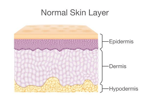
Layers of Skin: Epidermis, Dermis, Hypodermis
Structure of Skin
[Click Here for Sample Questions]
The structure of Skin can be explained in three parts:
Epidermis
The epidermis is the outermost layer of the skin. The cells present in the Epidermis include:
- Keratinocytes are made up of a protein namely keratin, the function of keratin is to strengthen the skin and make it waterproof. They are essentially considered skin cells.
- Melanocytes are cells that produce melanin. Melanin helps with the protection of skin from harmful sun rays. Melanin’s quantity in one’s body is dependent on the genes.
- Merkel Cells: Merkel cells have substances that often act as hormones. These cells are also quite close to the nerves.
- Langerhans cells: Epidermis also contains langerhans cells. Langerhans cells are specialised to protect the body from any foreign particles.
The various layers of the epidermis are:
- Stratum corneum
- Granular cell layer
- Spinous cell layer
- Basal cell layer
Dermis
Dermis is the part of the skin that has connective tissues. This layer is beneath the epidermis and has papillae, a substance that has finger-like projections. Other than papillae, this layer also has fat, collagen and fibres that make the skin flexible and strong. Dermis mainly functions to provide sensation, producing oil and sweats and also helps in the growth of hair.
The dermis can be further divided into two regions:
- Papillary region: This region has connective tissues which are loose and it also has finger-like projections, these projections push into the epidermis. Hence, giving the dermis a rather bumpy surface.
- Reticular region: This region is very dense. It has protein fibres present that contribute to elasticity as well as strength.
Hypodermis
Hypodermis is the deepest part of the skin. This part is made up of fat and is also called the subcutaneous layer. This storage of fat is crucial because it helps in regulating temperature and also provides soft cushion-like protection to the internal organs in the body.
The adipocyte cells present in the hypodermis have cells that store fat. They often act as energy reserves and also provide cushion-like protection to the organs and structures in the body in cases of trauma.
Function of Skin
[Click Here for Sample Questions]
Some of the functions of the skin include:
Sensation :- The most commonly known function of the skin is sensation. The skin helps us feel sensations such as pain, heat, cold, pressure. There is a network of nerves present in the skin sending signals to the brain.
Camouflage :- Some animal skins have camouflage properties that protect them from predators. Chameleons can release different pigments in their skin as per their will.
Temperature regulation :- Our body releases water through the skin by the process of perspiration. This helps in regulating the temperature of the body.
Storage :- The skin also stores fat and water in the tissues present. This helps with insulation.
Prevention of water loss:- The skin in the human body is thick which helps in preventing the loss of water in our body.
Excretion of scents :- Often animals use their scents as a way of marking their territory. This scent gives out a lot of information about their age, gender and often also the availability to their mate.
Protection of the environment :- Probably the most direct function of the skin is protecting the cells underneath from the external environment.
Also read: Difference between Cell and Tissue
Types of Tissue
[Click Here for Sample Questions]
The structure of the cells is dependent on the function it performs. The types of tissues present in the skin include
- Epithelial: Epithelial tissue is singularly referred to as epithelium. It has a free surface, facing either the body fluids or the outside environment. The cells present here are tightly packed with little intercellular matrices. The epithelial tissues can be further differentiated as simple and compound. The simple epithelium is further divided into squamous, cuboidal and columnar.

Epithelial tissues
- Connective Tissues: These tissues are the most widely present in the body of complex animals. They help in linking different tissues and organs in the body. The connective tissues range from soft connective tissues to specialised types. The connective tissue is of three types: loose, dense and specialised.
- Muscular Tissues: These muscle tissues are long and have a cylindrical fibre-like structure. These tissues play an active role in the overall movement in the body. Muscular tissues of three types are skeletal, smooth and cardiac.

Muscular Tissues
- Neural Tissues: These tissues have the greatest amount of control over the body's responsiveness to changes in conditions.

Neural Tissues
Things to Remember
- The skin has three major layers, that is, Epidermis, Dermis and Hypodermis.
- Some important components of these various layers of the skin include keratinocytes, Melanocytes, Langerhans cells and Merkel cells.
- There are four types of tissues in the body, that is, connective, muscular, neural and epithelial.
- Some important functions of skin include sensation temperature regulation, protection of organs, storage, excretion of scents and prevention of water loss.
- The epidermis also has various layers within it which include the Stratum corneum, the Granular cell layer, Spinous cell layer and Basal cell layer.
Also read: Animal Tissues
Sample Questions
Ques. Explain the structure of the layers of the epidermis. (5 Marks)
Ans. The structure of layers of the epidermis has the following elements:
- Stratum Basale is the deepest epidermal layer and attaches its epidermis to the basal lamina, a layer below the dermis. This layer is primarily made up of basal cells which essentially stem cells that have a cuboidal structure to them. Basal cells further can be divided into Merkel cells and melanocytes. The former functions as a receptor and is important for stimulating sensory nerves and the latter produces a pigment called melanin.
- Stratum Spinosum has over eight to ten layers of keratinocytes. Another cell present in this layer is the Langerhans cells which are a type of dendritic cell that consumes bacteria and foreign materials present in the layer.
- Stratum Granulosum is grainy in its appearance because of the changes due to the presence of keratinocytes being pushed from stratum spinosum.
- Stratum lucidum is a translucent layer above the stratum granulosum and below the stratum corneum. Here the keratinocytes area dead and are densely packed with Leiden- a protein that is clean and also rich in
- Stratum Corneum is a superficial layer of the epidermis that is also exposed to the outside environment. Around 15-30 layers of cells are present in this layer. This layer is then replaced constantly in four weeks.
Ques. Explain the structure of the layers of the dermis. (3 Marks)
Ans. The papillary layer is made of loose, areolar connective tissue, which means the collagen and elastin fibres of this layer form a loose mesh. This superficial layer of the dermis projects into the stratum basale of the epidermis to form finger-like dermal papillae.
Reticular Layer is made of dense and irregular connective tissue. It is well vascularized and has a rich sympathetic nerve supply. It is a thicker reticular layer. It also has Elastin fibres which give some elasticity to the skin.
Ques. Describe hypodermis and the deep fascia. (3 Marks)
Ans. The hypodermis, also known as the subcutaneous tissue, rests above the deep fascia which is a deep connective tissue surrounding the individual or group muscles that separates into different fascial compartments. It is a discrete fibrous layer.
Ques. What is the function of keratinocytes? (3 Marks)
Ans. The functions of Keratinocytes are as follows:
- Keratinocyte is a fibrous protein due to which hair, nails and skin have hardness and also have water-resistant properties.
- Keratinocytes have an essential role in skin repair, they help in the re-epithelialization process.
- It also forms a barrier to prevent damage to the skin.
Ques. What is the function of Melanocytes? (3 Marks)
Ans. The functions of Melanocytes are as follows:
- Melanocytes help in the production of melanin and affect the colour of the skin.
- Dendrites of melanocytes are key for the transfer of melanin to the adjacent epidermal cells.
- Melanocytes and Keratinocytes together function to form the epidermal-melanin unit.
Ques. Explain the anatomy of the different layers of the skin. (3 Marks)
Ans. The skin has various elements and substances which comprises pigmentation related cells such as melanin which is formed by melanocytes. It also has mesodermal cells. Further, the substances in the skin also have enzymes that help in repairing DNA, it does the same by reversing the UV damage. In case this enzyme is absent, it increases the chances of skin cancer in humans.
Ques. Differentiate between Papillary Region and Reticular Region. (3 Marks)
Ans. The difference between Papillary Region and Reticular Region are:
| Papillary Region | Reticular Region |
|---|---|
| It is made of loose connective tissue. | It is made of dense and irregular connective tissues. |
| It has finger-like projections which push in the epidermis. | It also has protein fibres in the reticular regions. |
| It provides nutrient-related supply and also regulates the temperature as well. | It provides skin strength and also elasticity. |
| The papillary region lies below the epidermis. | It is below the papillary layer. |
| It is very highly vascularised. | It is less vascularised. |
| Some of the cells in the papillary region include mast cells, macrophages and leukocytes. | This layer has very fewer cells than the papillary layer. |
Ques. Differentiate between Epidermis and Dermis. (3 Marks)
Ans. The difference between Epidermis and Dermis are as follows:
| Epidermis | Dermis |
|---|---|
| It is a thick layer that has living cells below the epidermis containing blood vessels and sweat glands. | It is the outer layer of the cell and protects and faces the body from the external environment. |
| It is most often only found in animals. | It is found in both plants and animals. |
| It has an extracellular matrix. | It is very tightly packed without the presence of an extracellular matrix. |
| It gets oxygen and also has nutrients from blood capillaries. | It gets oxygen and nutrients from diffusion from the dermis. |
| It also has sensory nerve endings. | It does not have any nerves. |
| It is composed of living cells. | It is composed of both living and non-living cells. |
Ques. Differentiate between Epithelial tissues and Connective tissues. (3 Marks)
Ans. The difference between Epithelial tissues and Connective tissues are as follows:
| Epithelial tissues | Connective tissues |
|---|---|
| The cells in Epithelial tissues are close together. | The connective tissues have very scattered cells. (extracellular matrix). |
| It acts as a barrier for microorganisms and dehydration when secretion, excretion and absorption of substances happen. | It supports and also helps in connecting tissues and organs as well. |
| It is found over the basal membrane. | It is found underneath the basal membrane. |
| There are no blood capillaries in this tissue. | It has a lot of blood capillaries that absorb nutrients. |
| It lines the cavity and surfaces of organs and blood vessels. | It separates and connects different types of tissues and organs in the body. |
| It is commonly found in glands, organs like kidneys and lungs. | It is commonly found in adipose, cartilage and muscles. |
Ques. Differentiate between Muscular tissues and Neural tissues. (3 Marks)
Ans. The difference between Muscular tissues and Neural tissues are as follows:
| Muscular tissues | Neural tissues |
|---|---|
| It specialises in contraction. | It specialises in communication. |
| It is a specialised animal tissue for contraction. | It is a specialized animal tissue for communication. |
| It functions in smooth, cardiac and skeletal muscle. | It functions in the brain and spinal cord. |
| It originates from mesoderms. | It originates from ectoderm. |
| It consists of fibres, sarcomeres. | It consists of the cytoplasmic process and the cell body. |
| It comprises a lot of myofibrils. | It consists of a lot of nerve cells. |
Check-Out:


Comments